
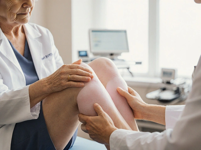
Oedema is a medical condition characterized by excess fluid accumulation in the interstitial spaces of the body, commonly presenting as swelling in the legs, ankles, or hands. It can arise from vascular, cardiac, renal, or lymphatic dysfunction.
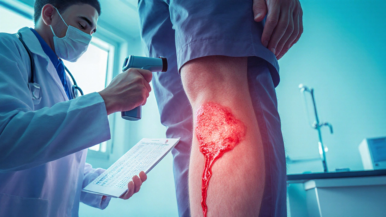
Cellulitis is a bacterial infection of the skin and subcutaneous tissue that leads to redness, warmth, and painful swelling. The most frequent pathogens are Streptococcus pyogenes and Staphylococcus aureus.
What’s the difference between oedema and cellulitis?
At a glance both conditions cause swelling, but the underlying mechanisms diverge. Oedema stems from fluid leakage or impaired drainage, whereas cellulitis results from an active infection that triggers inflammation. Recognizing this distinction guides appropriate treatment - diuretics for fluid overload versus antibiotics for bacterial invasion.
Key causes
Both entities share some risk factors but also have unique triggers.
- Venous insufficiency - faulty veins in the lower limbs increase hydrostatic pressure, a classic cause of chronic oedema.
- Lymphatic obstruction - blockage of lymph channels, often after surgery or radiation, leads to lymphedema.
- Heart failure - reduced cardiac output raises venous pressure, spilling fluid into tissues.
- Kidney disease - impaired sodium excretion retains water, swelling the extremities.
- Bacterial infection - breaks in the skin allow bacteria to invade, sparking cellulitis.
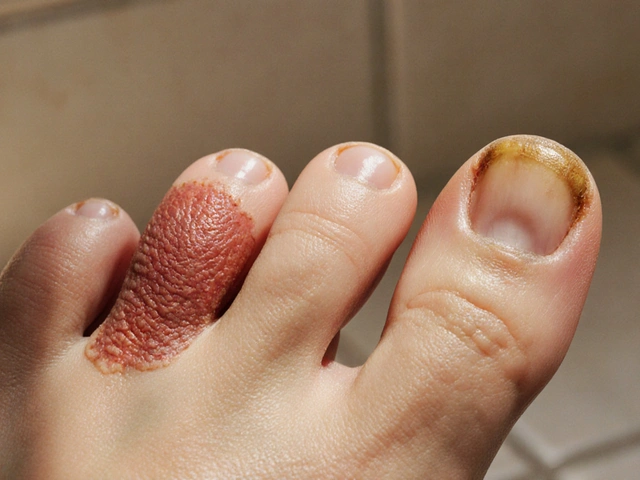
- Skin conditions such as athlete’s foot or eczema create portals for microbes.
Typical symptoms
Understanding the clinical picture helps you decide whether you’re looking at oedema or cellulitis.
| Feature | Oedema | Cellulitis |
|---|---|---|
| Onset | Gradual, often over days or weeks | Rapid, within hours |
| Skin colour | Pale or slightly shiny | Red to deep pink, may spread |
| Temperature | Usually normal | Warm or hot to touch |
| Pain | Mild discomfort, heaviness | Sharp, throbbing pain |
| Systemic signs | Rare | Fever, chills possible |
How doctors diagnose the two
Physical exam remains the cornerstone. Clinicians look for pitting on pressure (a hallmark of oedema) versus raised, tender borders (typical for cellulitis). Imaging such as Doppler ultrasound can rule out deep vein thrombosis, a frequent mimic. Laboratory tests - a full blood count may reveal elevated white cells in cellulitis, while serum albumin helps gauge oncotic pressure‑related oedema.
Treatment options
Therapeutic goals differ, so a one‑size‑fits‑all approach won’t work.
Managing oedema
- Diuretics - medications like furosemide promote renal excretion of water and sodium, shrinking excess fluid. Dosage must be tailored to kidney function.
- Compression therapy - graduated stockings apply external pressure, encouraging venous return and lymph drainage.
- Elevation of the affected limb above heart level for 15‑30 minutes several times a day.
- Low‑salt diet and adequate protein intake to support oncotic balance.
- Physical therapy focusing on calf‑muscle pump activation.
Managing cellulitis
- Antibiotics - empirical oral agents such as cephalexin or clindamycin cover common streptococcal and staphylococcal species. Intravenous therapy is reserved for severe cases or when oral absorption is doubtful.
- Analgesics like ibuprofen reduce pain and inflammation.
- Wound care: gentle cleansing, sterile dressings, and monitoring for abscess formation.
- Addressing underlying portal of entry - treating athlete’s foot, improving skin hygiene.
When to seek urgent care
If swelling spreads rapidly, is accompanied by high fever (>38.5°C), or you notice spreading redness that crosses joint lines, call emergency services. These signs may indicate a systemic infection or septicemia, especially in immunocompromised patients.
Prevention strategies
Pre‑emptive steps can curb both conditions.
- Maintain a healthy weight - excess adipose tissue raises venous pressure.
- Exercise regularly to boost circulation; simple calf raises are effective.
- Inspect feet daily if you have diabetes; treat any cuts promptly.
- Avoid prolonged standing or sitting without movement; take a 5‑minute walk every hour.
- For patients with known venous insufficiency, wear properly fitted compression garments.
Related conditions and next topics to explore
Understanding oedema and cellulitis opens doors to a wider network of vascular and dermatologic issues. Readers often want to learn about lymphedema management, deep vein thrombosis, and chronic venous ulcer care. Future articles will dissect the role of hormonal fluctuations in peripheral swelling and review new oral antibiotics for resistant cellulitis strains.
Conclusion
Both oedema and cellulitis present with swelling, but the former is a fluid‑balance problem while the latter is an infection. Accurate identification steers treatment toward diuretics or antibiotics, respectively, and prevents complications. Keep an eye on symptom evolution, and don’t hesitate to involve a healthcare professional when the picture becomes unclear.
Frequently Asked Questions
Can oedema turn into cellulitis?
Yes, if the swollen skin cracks or a wound forms, bacteria can enter and cause cellulitis. Prompt skin care and hygiene reduce this risk.
Are over‑the‑counter creams effective for cellulitis?
Topical creams alone cannot treat the deeper infection of cellulitis. They may soothe irritation, but systemic antibiotics are required to clear the bacteria.
What lifestyle changes help reduce chronic oedema?
Weight management, regular leg elevation, low‑salt diet, compression stockings, and daily calf‑muscle exercises are the core strategies recommended by NICE and the American Heart Association.
How long does antibiotic treatment for cellulitis usually last?
For uncomplicated cases, a 5‑7day course of oral antibiotics is standard. Severe or resistant infections may need 10‑14days of intravenous therapy.
Is swelling in the arms ever a sign of cellulitis?
Although less common than leg involvement, cellulitis can affect the arms, especially after an injury or intravenous line. Look for redness, warmth, and rapid progression.


janvi patel
September 26, 2025 AT 18:01Oedema isn’t always as harmless as the article suggests.
Lynn Kline
September 29, 2025 AT 01:35Fantastic job breaking this down!!! Your guide is both stellar and insightful!!! Keep the amazing content flowing!!!
Rin Jan
October 2, 2025 AT 12:55Reading through this piece reminded me just how crucial it is to distinguish fluid overload from an active infection because mistaking one for the other can lead to inappropriate therapy and potentially dangerous outcomes. While oedema may present as a painless, gradual swelling doctors often rely on pitting edema tests and serum albumin levels to confirm a fluid shift rather than an inflammatory response. In contrast cellulitis erupts quickly with warmth, redness and systemic signs such as fever that demand prompt antimicrobial coverage. Ignoring these temporal patterns can result in delayed antibiotics and worsening sepsis. Moreover the underlying etiologies differ; heart failure and renal disease drive hydrostatic pressure while bacterial invasion exploits skin breaches. Compression therapy and diuretics have no place in treating a bacterial infection and may even mask its progression. Conversely unnecessary antibiotics for simple oedema contribute to resistance and expose patients to needless side effects. The article correctly emphasizes elevation and low‑salt diets for chronic fluid retention but could have highlighted the role of regular monitoring of weight and leg circumference as objective markers. It also briefly mentioned lymphatic obstruction yet omitted the importance of manual lymphatic drainage techniques that many physiotherapists employ. Lastly patient education on skin hygiene cannot be overstated because even minor fissures can become portals for staphylococcal entry turning a benign edema into a fulminant cellulitis. In summary the distinction is not merely academic it directly informs the therapeutic pathway and ultimately patient safety.
Jessica Taranto
October 4, 2025 AT 06:35I appreciate how the guide lays out both conditions side‑by‑side; the table makes it easy to spot the rapid onset and warmth that flag cellulitis versus the slow, painless nature of oedema. It’s a solid reference for anyone trying to self‑monitor symptoms.
akash chaudhary
October 7, 2025 AT 04:01The article glosses over critical diagnostic work‑ups; relying on “full blood count may reveal elevated white cells” is lazy when a CRP, ESR and wound culture are standard for cellulitis assessment. Also, the suggestion that compression can be used indiscriminately ignores contraindications such as acute infection or arterial insufficiency. Readers need a stricter protocol, not vague generalities.
Adele Joablife
October 9, 2025 AT 11:35While the guide is thorough, it could benefit from highlighting that not all patients tolerate diuretics and that electrolyte monitoring is essential. A balanced view on potential side‑effects would make the piece more complete.
kenneth strachan
October 11, 2025 AT 13:35Honestly, the whole thing feels a bit too clinical-where’s the real‑life story of someone who mistook oedema for cellulitis and ended up in the ER? Add some drama next time, it’ll be more relatable!!!
Mandy Mehalko
October 13, 2025 AT 12:48Great info! I feel more confident now to check my own legs and know when to call a doc. Keep the positivity coming!!!
Bryan Kopp
October 15, 2025 AT 06:28Not all oedema is benign.
Patrick Vande Ven
October 17, 2025 AT 14:01In response to the extensive commentary above, it is worth noting that the differentiation between oedema and cellulitis hinges on both clinical presentation and targeted investigations. A methodical approach that incorporates Doppler ultrasound to exclude deep vein thrombosis, alongside inflammatory markers, enhances diagnostic accuracy. Moreover, treatment algorithms should reflect patient‑specific factors such as comorbid heart failure or diabetes, ensuring that diuretic therapy is not administered in the presence of an active infection without appropriate antimicrobial coverage.
Tim Giles
October 18, 2025 AT 23:21Building on the formal suggestions provided, clinicians must also remain vigilant for atypical presentations; for instance, cellulitis can occasionally manifest without pronounced erythema in immunocompromised individuals, making reliance on temperature and pain cues indispensable. Additionally, the role of serial measurements-both of limb circumference and weight trends-cannot be overstated in tracking oedema resolution, particularly when patients are on long‑term diuretic regimens. Education on proper compression garment fitting is another cornerstone that prevents iatrogenic injury, especially in those with concurrent arterial disease where excessive pressure may precipitate ischemia. Finally, interdisciplinary collaboration with physiotherapists for manual lymphatic drainage complements medical therapy, offering a holistic strategy that addresses both fluid dynamics and tissue health.
Peter Jones
October 20, 2025 AT 11:28Love how the community is adding layers to the discussion; the practical tips on limb measurement and compression fitting really round out the guide. Thanks for the collaborative spirit!
Gerard Parker
October 22, 2025 AT 19:01To sum up, accurate differentiation between oedema and cellulitis is essential for safe patient care. Use physical exam findings, appropriate imaging, and lab work to guide therapy-diuretics for fluid overload, antibiotics for infection. Stay vigilant for mixed presentations and adjust treatment plans accordingly.